
In 2024, we introduced you to Sydney “Syd” Sattler, then a PhD candidate in the Abitua Lab in UW’s Department of Genome Sciences, as the first UW recipient of the Shurl and Kay Curci Foundation PhD Fellowship. At the time, she was just beginning to chase a question about how immune cells decide what to become and when.
Two years later, that question has an answer, and it’s stranger and more interesting than expected.
The Findings
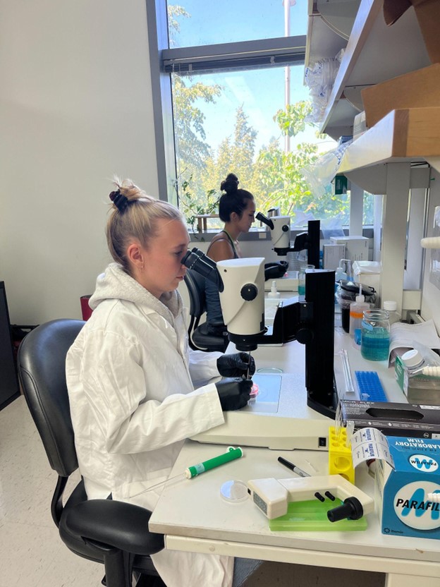
Sattler and her colleagues in the Abitua Lab working alongside Nicholas Lammers, Sebastian Hriscu, Clare Booth, Cole Trapnell, and PI Philip Abitua have been studying the annual killifish, Nothobranchius furzeri, a small fish whose embryos develop on an unusually compressed and altered timeline.
Annual killifish inhabit temporary pools that form during seasonal rains and disappear during dry periods. Adult fish survive long enough to reproduce and deposit embryos into the substrate before the pools evaporate. The embryos persist in the dry sediment until favorable conditions return. Adaptation to this unpredictable environment has produced extensive evolutionary changes in lifespan and early embryonic development. Unlike other vertebrates’ models, the embryonic cells of annual killifish disperse during early development before later reaggregating to form the embryonic axis.
In every vertebrate studied so far, the body follows a predictable order of operations during early development. Gastrulation, the stage where an embryo’s cells organize into the layers that will become different tissues happens first. Only after that do primitive immune cells, like neutrophils, emerge from a specific tissue layer (ventral mesoderm) that gastrulation creates.
The team found that in killifish, that order breaks. Neutrophils show up before gastrulation even happens; before the tissue, they’re “supposed” to come from has even formed. The blood vessel–forming cells still show up later, on schedule, from that same tissue. But the immune cells jumped off the line.
Using a combination of imaging, single-cell sequencing, and signaling experiments, the team showed this isn’t a fluke: blocking a specific signaling pathway (BMP) shuts down the early immune cell program, while blocking the pathway that normally drives gastrulation (Nodal) doesn’t touch it. In other words, the cells are running on the same genetic circuitry immune cells always use; it’s just been unplugged from its usual timing and wired earlier.
Why it matters: It’s a clear example of how evolution can take a conserved, reliable genetic program and redeploy it, new timing, new location, same underlying tool, when a species’ whole embryonic architecture shifts. For a field trying to understand how immune systems form (and sometimes malform) across species, including our own, it’s a reminder that developmental “rules” are more like defaults than laws.
The Person Behind the Preprint; Sattler’s Perspective:
As science typically goes, there wasn’t one moment when Sattler and team realized that immune cells develop early in killifish. Instead, multiple findings kept adding up to a moment of conviction. The team first saw a subset of cells that moved rapidly during early development and recognized that many immune cell types are known to move at high speeds. Next, they were able to show that a subset of cells expressed genes that are known markers of immune cells using single-cell RNA sequencing, but they couldn’t be sure these were the same rapidly moving cells. The team was not fully convinced until they could put these two observations together. Using CRISPR, Sattler integrated a green fluorescent protein next to L-plastin, a gene that is known to only be expressed in immune cells. This tool allowed Sattler to highlight immune cells live in the killifish embryo, confirming that the same subset of cells that were first seen moving rapidly prior to gastrulation were indeed the population that was expressing immune markers.
“I’ll never forget the first movie I took of the immune cells, lit up green, flitting around in the early embryo. I must have stared at that movie for 20 minutes straight, because seeing really is believing. “
Another big turning point in the project was when the team was able to computationally track individual immune cells back in time to their origins in the embryo. Many of the experiments aimed at origin were performed on fixed tissue, meaning they were only able to glimpse a snapshot of where the cells were in the embryo at a given time.
Cell tracking allowed Sattler to follow a cell through its developmental history to see the moment when it became an immune cell. This analysis showed that the immune progenitor cells began to first move more rapidly compared to other embryonic cells, then slow down, divide, and produce two daughter cells that then differentiated into immune cells. They were able to see that this process happened dynamically over many hours at the periphery the aggregate; where the embryo begins to form.
Researchers were always asking where these cells came from, and after this moment Sattler finally had a strong answer for them.
What’s Next
The paper is currently under revision for journal submission; the preprint represents where the science stands today, with peer review still ahead.
Sattler expects to complete her PhD this Fall and is looking toward an industry role focused on adaptive immune cells and their role in autoimmune disease, a natural next chapter for someone who’s spent the last several years figuring out how immune cells get their start in the first place.